Elkhart Clinic Imaging Services
The Elkhart Clinic Imaging department performs approximately 18,000 exams a year that includes CT, Dexa, MRI, PET, Ultrasound, and
X-ray. These services are provided by our Imaging staff that consists of nurses, schedulers and technologists. All of our staff are trained professionals and are licensed and certified in their fields. Imaging services at the clinic are broken into three separate departments: Advanced Imaging, Imaging and Surgery.
Advanced Imaging is housed in our new state of the art facility that was added on to the main building in 2013. Advanced Imaging has it’s own separate entrance on the West side of the main building. Patients use this entrance to register for the following exams: CT, MRI, and PET/CT imaging. All Advanced Imaging studies are scheduled exams and you should call (574) 296-3480 to schedule an appointment or to talk to someone about these exams. These modalities are all accredited by the American College of Radiology (ACR) and hold current state licenses.

CT (Computed Tomography)
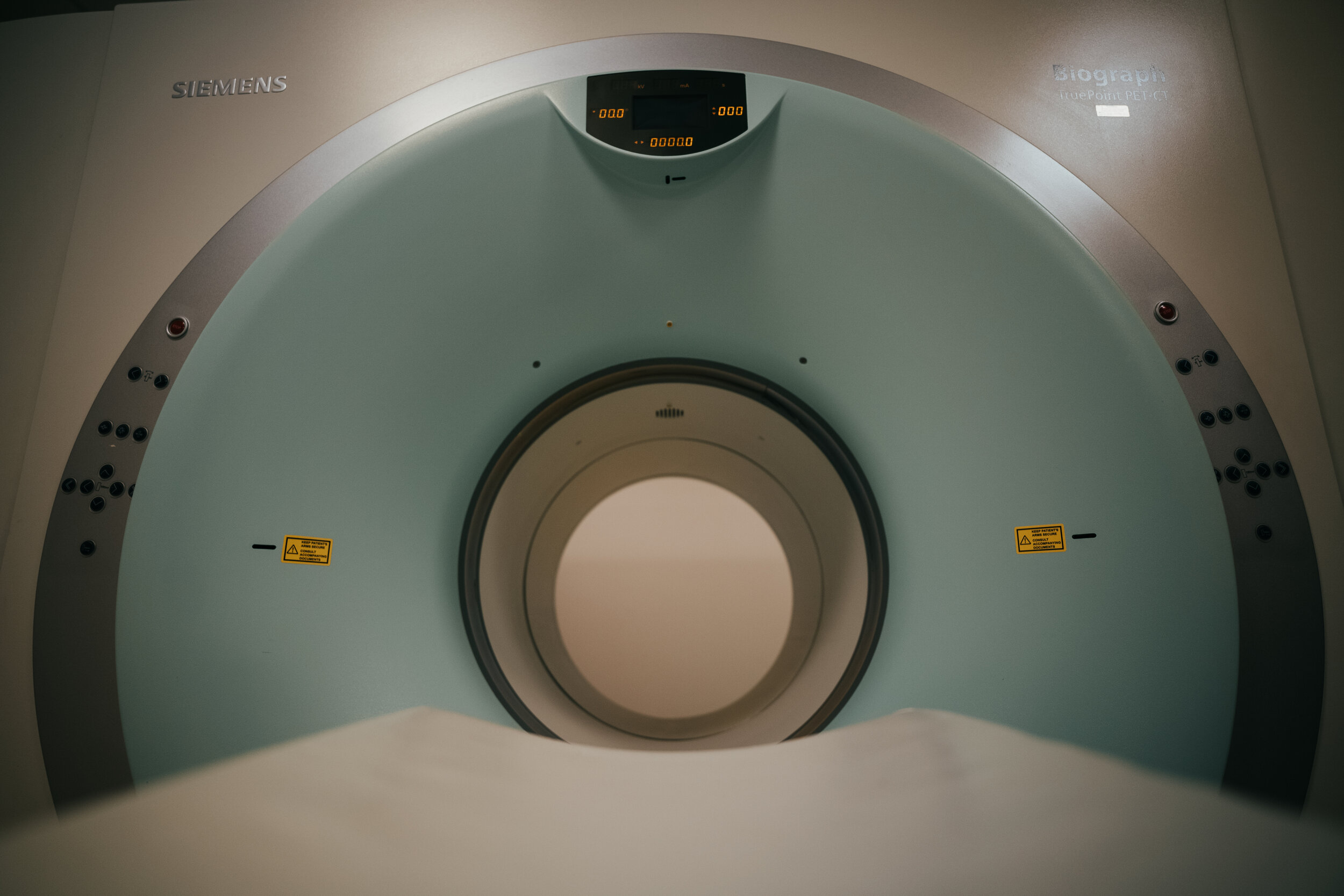
CT imaging uses radiation to produce cross-sectional images or “slices” of anatomy, like the slices in a loaf of bread. The cross-sectional images are used for a variety of diagnostic and therapeutic purposes. Computed Tomography is used to detect bone fractures, cancer, lung disease, stroke, head bleeds, kidney stones, appendix, liver disease, urinary tract problems, aneurysms, vessel stenosis, bowel/stomach issues, and much more. Patients are put onto a table that slides back and forth through a “donut” shaped machine, and most exams only last a few minutes. Claustrophobia is usually not an issue due to the speed of the test and the size of the machine. Some exams require contrast to be administered depending on the exam and the diagnosis. The contrast can be given orally, IV or sometimes via both routes.
MRI (Magnetic Resonance Imaging)
MRI is a radiology procedure that uses magnetism, radio frequencies, and a computer to produce images of body structures. It is used to diagnose many different problems or issues. Some of those issues are: Cancer, MS, tears in ligaments/tendons/muscles, bleeds, strokes, TIA’s, aneurysms, lesions, masses, stress fractures, infections, spinal stenosis, sciatica, and ruptured discs. Usually we scan one body part at a time, due to the long scan times. Each scan ranges from 30 minutes up to about an hour for one body part. The patient is placed on a table and is moved into the MRI machine which resembles a tunnel. The body part being scanned is placed in a coil which helps capture the radio waves and helps create the image; then placed in the center of the tunnel. Claustrophobia is a concern in MRI; for those patients who experience nervousness in closed spaces we suggest they ask their physician for some medicine to relax them. Our MRI technologists are trained to work with claustrophobic patients to assist them during the exam. MRI machines make loud knocking and vibration noises, and ear protection is given to all patients. Contrast is given to patients for some exams depending on body part being scanned and the accompanying diagnosis.
PET/CT (Positron Emission Tomography)
PET/CT is a type of Nuclear Medicine exam that uses small amounts of radioactive material to diagnose, treat, and determine the severity of a variety of diseases. One of the main uses of PET/CT is to help stage cancer; but it is also used for other diseases throughout to the body. Because PET/CT is used to pinpoint molecular activity in the body, it offers the potential to identify diseases at their earliest stages and also determines the patient’s immediate response to therapeutic interventions. PET/CT exams take about 2 hours for the the entire exam process. A radioactive tracer is injected into the patient, and then a patient relaxes in a room by themselves for an hour. The patient is then placed on a table and are moved through a tunnel. During the scan portion of this test; a set of CT images are taken and then a set of PET images are taken. The two sets of images are then combined to get a detailed cross sectional image that helps locate any potential disease.
Imaging is located in the main building and patients register at the main entrance for the following exams: Dexascan, Ultrasound, and X-ray. Our schedulers and technologists are all trained and licensed professionals that work extremely hard to provide quality and friendly services to all our patients. Patients can call (574) 296-3331 to schedule an appointment or to talk with someone about these services. Our services are licensed by the state and accredited by the Intersocietal Accreditation Committee (ICAVL).
Dexascan (Dual Energy X-ray Absorptiometry)
This is the preferred technique when it comes to measuring bone mineral density. Dexa is a machine that produces two X-ray beams, each with different energy levels. One beam is high energy and the other beam is low energy, and the amount of X-rays that pass through the bone is measured for each beam. Based on the difference between the two energy levels determines the density of the bone. In general, Dexa’s produce very low amounts of radiation exposure; and are typically done on the hip or spine. Dexa scans last between 10 and 20 minutes and are non-invasive.
Ultrasound
A Medical test that uses high-frequency sound waves to capture live images from the inside of your body. For this reason, physicians usually like to use this modality on pregnant patients to help diagnose problems, due to the fact no radiation is involved and does not harm the fetus. This modality also allows for your doctor to see problems with your organs, tissues and vessels without having to make any incisions into your body. It is also used during surgeries, biopsies and pain injections to help guide the needle to the proper location; and can be used to help determine vessel stenosis and blood flow.
X-ray
Used in the identification, diagnosis and treatment of many medical conditions; X-ray is a way that a physician can look into your body in an easy and painless way. It uses x-ray beams (small amounts of radiation) to look at bones, lungs, joints, foreign bodies, etc. Physicians use it to diagnose fractures, dislocations, infections, lung disease/issues, locations of foreign bodies, arthritis, and many other problems that a patient may be experiencing. Usually an X-ray is the very first test that is done on a patient that is experiencing pain, swelling, coughing, shortness of breath, bumps, limited mobility, lack of strength, trauma, etc. An x-ray can be taken of any body part.
Surgery imaging is done at our surgery center with a machine called a C-arm. This device is basically used during surgeries to help aid physicians in the surgery process. Specialists in fields such as surgery, orthopedics, trauma, vascular surgery and cardiology use C-arms for intraoperative imaging. This device provides high resolution x-ray images in real time (Fluoro Imaging) so physicians can make changes as needed during the surgery and ultimately help recovery time, eliminate follow-up surgeries and reduce cost to the patient and healthcare facility.


